- Anatomy
- Conditions
- Procedures

Knee Pain
Knee pain is a common condition affecting individuals of various age groups. It not only affects movement but also impacts your quality of life. An injury or disease of the knee joint or any structure surrounding the knee can result in knee pain. A precise diagnosis of the underlying cause is important to develop an appropriate treatment plan.

Knee Injury
Pain, swelling, and stiffness are the common symptoms of any damage or injury to the knee. If care is not taken during the initial phases of injury, it may lead to joint damage, which may end up destroying your knee.

Knee Sports Injuries
Trauma is any injury caused during physical activity, motor vehicle accidents, electric shock, or other activities. Sports trauma or sports injuries refer to injuries caused while playing indoor or outdoor sports and exercising. Sports trauma can result from accidents, inadequate training, improper use of protective devices, or insufficient stretching or warm-up exercises. The most common sports injuries are sprains and strains, fractures, and dislocations.

Anterior Knee Pain
Anterior knee pain is characterized by chronic pain over the front and centre of the knee joint. It is common in athletes, active adolescents (especially girls) and overweight individuals. Anterior knee pain refers to various conditions, which include runner's knee or patellar tendinitis, and chondromalacia of the patella. There is an inter-individual variation in the duration and presentation of pain.

Runner's Knee
Patellofemoral pain syndrome also called runner’s knee refers to pain under and around your kneecap. Patellofemoral pain is associated with a number of medical conditions such as anterior knee pain syndrome, patellofemoral malalignment, and chondromalacia patella. Patellofemoral pain is a common complaint among runners, jumpers, and other athletes such as skiers, cyclists, and soccer players; thus the common name, runner’s knee.

Jumper's Knee
Jumper’s knee, also known as patellar tendinitis, is inflammation of the patellar tendon that connects your kneecap (patella) to your shinbone. This tendon helps in the extension of the lower leg. Jumper’s knee usually results from repetitive trauma or overuse, particularly from sports activities that involve jumping such as basketball or volleyball.

Kneecap Bursitis
Bursitis refers to the inflammation and swelling of the bursa. Inflammation of the bursa in front of the kneecap (patella) is known as kneecap bursitis or prepatellar bursitis. Symptoms of kneecap bursitis include pain and swelling in front of the knee. You may also experience tenderness, warmth, and redness on the front of the knee.

Patellar Instability
Any damage to the supporting ligaments may cause the patella to slip out of the groove either partially (subluxation) or completely (dislocation). This misalignment can damage the underlying soft structures such as muscles and ligaments that hold the kneecap in place. Once damaged, these soft structures are unable to keep the patella (kneecap) in position.
Patellofemoral Instability
Patellofemoral instability means that the patella (kneecap) moves out of its normal pattern of alignment. This malalignment can damage the underlying soft structures such as muscles and ligaments that hold the knee in place. Patellofemoral instability can be caused because of variations in the shape of the patella or its trochlear groove as the knee bends and straightens.

Lateral Patellar Instability
Lateral patellar instability is defined as a lateral shift or displacement of the patella (kneecap) as a result of disruptive changes in the medial patellofemoral ligament (MPFL) and medial patellar retinaculum. Lateral patellar instability can be caused by variations in the shape of the patella or its femoral groove as the knee bends and straightens. Normally, the patella moves up and down within the femoral groove when the knee is bent or straightened.

Medial Patellar Instability
Medial patellar instability is a disabling condition characterised by medial subluxation of the patella which occurs as a complication of lateral retinacular release surgery. Lateral retinacular release is a surgical procedure employed for the treatment of lateral patellar instability. However, disruption of the lateral patellar structures during this surgery has been associated with medial patellar instability.

Patellar Dislocation/Patellofemoral Dislocation
Patellar dislocation occurs when the patella moves out of the patellofemoral groove, (trochlea) onto the bony head of the femur. If the kneecap partially comes out of the groove, it is called subluxation; if the kneecap completely comes out, it is called dislocation (luxation).
Patellar Tendon Rupture
The patellar tendon works together with the quadriceps muscle and the quadriceps tendon to allow your knee to straighten out. Patella tendon rupture is the rupture of the tendon that connects the patella (kneecap) to the top portion of the tibia (shinbone).

Patella Fracture
The kneecap or patella forms a part of the knee joint. It is present at the front of the knee, protecting the knee and providing attachment to various muscle groups of the thigh and leg. The undersurface of the kneecap and the lower end of the femur are coated with articular cartilage, which helps in smooth movement of the knee joint. A fracture in the kneecap is rare but common in adult males.

Fractures of the Patella
The patella or kneecap is a small bone present in the front of your knee where the thigh bone meets the shinbone. It provides protection to your knee and attachment to muscles in the front of the thigh. An injury to the knee can result in a break or fracture of the patella.

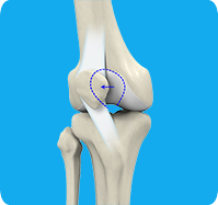
Osteochondritis Dissecans of the Knee
Osteochondritis dissecans is a joint condition in which a piece of cartilage, along with a thin layer of the bone separates from the end of the bone because of inadequate blood supply. The separated fragments are sometimes called “joint mice”. These fragments may be localized or may detach and fall into the joint space, causing pain and joint instability.

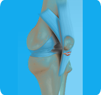
Osteochondral Defect of the Knee
An osteochondral defect, also commonly known as osteochondritis dissecans, of the knee refers to a damage or injury to the smooth articular cartilage surrounding the knee joint and the bone underneath the cartilage. The degree of damage may range from a rupture of the cartilage to a slight crack of the bone to a piece of the bone breaking off within the joint.

Quadriceps Tendon Rupture
The quadriceps tendon is a thick tissue located at the top of the kneecap. It works together with the quadriceps muscles to allow us to straighten our leg. The quadriceps muscles are the muscles located in front of the thigh. The quadriceps can rupture after a fall, direct blow to the leg and when you land on your leg awkwardly from a jump. Quadriceps tendon rupture most commonly occurs in middle-aged people who participate in sports that involve jumping and running.

Iliotibial Band Syndrome
Iliotibial band syndrome is an overuse injury resulting from the inflammation of the iliotibial band. It occurs when the iliotibial band and the lower outside portion of the thighbone at the knee joint rub against each other. Iliotibial band syndrome commonly occurs in athletes, cyclists, and runners, and can occur from quickly increasing distances with running or biking activities.

Knee Dislocation
Knee dislocation is a condition that occurs when the bones that form the knee joint, namely the femur or thigh bone get separated from the shin bone. This can cause serious damage to the nerves, blood vessels, and ligaments surrounding the knee, leading to a decline in strength and overall health of the leg.

Knee Fracture
A fracture is a condition in which there is a break in the continuity of the bone. In younger individuals, these fractures are caused by high energy injuries, as from a motor vehicle accident. In older people, the most common cause is a weak and fragile bone.

Knee Stress Fractures
Stress fractures of the patella or knee are very rare. Approximately two out of 10,000 athletes may experience a patella stress fracture. Initial symptoms include activity-related pain and then a fatigue stress fracture after minor trauma. The term insufficiency stress fracture is used for cases where the patella is weakened previously such as after patella resurfacing surgery.

Distal Femur Fracture
Distal femur fractures may be caused by high energy injuries such as a fall from a height or a motor vehicle accident. Patients with osteoporosis, bone tumours or infections, or a history of knee replacement are more prone to femur fractures. In the elderly, even a simple fall from a standing position may result in a fracture as the bones tend to become weak and fragile with advancing age.

Fractures of the Tibia
Fractures of the tibia vary depending on the force involved and are classified based on the location of the fracture, the pattern of the fracture, and exposure of the fracture site. The lower leg is made up of two long bones called the tibia and fibula that extend between the knee and ankle. The tibia or shinbone is the larger of the two bones. It bears most of the body’s weight and helps form the ankle joint and knee joint.
Stress Fracture of the Tibia
A stress fracture of the tibia or shinbone is a thin fracture, also called a hairline fracture that occurs in the tibia due to excess stress or overuse. The tibia is a weight-bearing bone in which stresses can accumulate from activities such as running and jumping.
Quadriceps Tendon Rupture and Repair
Quadriceps tendon rupture is an uncommon injury that usually warrants surgical treatment. Middle-aged individuals who are active in sports are more prone to having a quadriceps tendon rupture. With suitable and timely care, most individuals are able to return to their normal activities and occupations. Recovery varies from individual to individual and depends on active participation in your post-surgical rehabilitation exercises.

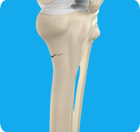
Tibial Eminence Fractures
The tibial eminence, also called the tibial spine, is a bony protuberance of the tibia (shin bone) that attaches to the anterior cruciate ligament (ACL) of the knee joint. A tibial eminence fracture is break or crack in the bony attachment of the ACL to the tibia. The fracture can be a contact or non-contact injury and occurs at the base of the tibial eminence.

Periprosthetic Knee Fractures
Knee replacement, also called knee arthroplasty, is a surgical procedure in which the worn-out or damaged surfaces of the knee joint are removed and replaced with artificial implants. Any resulting fractures or breaks in the bone around the implant are called periprosthetic knee fractures. These fractures may occur during surgery (intraoperative) or after surgery (postoperative), and usually involve the patella, tibia or the femur (kneecap, shinbone, and thighbone). Women are at higher risk than men.

Knee Sprain
Knee sprain is a common injury that occurs from overstretching of the ligaments that support the knee joint. A knee sprain occurs when the knee ligaments are twisted or turned beyond its normal range, causing the ligaments to tear.

Knee Ligament Injuries
The knee is a hinge joint made up of two bones, the thighbone (femur) and shinbone (tibia). Ligaments are tough bands of tissue that connect one bone to another bone. The ligaments of the knee stabilize the knee joint. There are two important groups of ligaments that hold the bones of the knee joint together, collateral and cruciate ligaments - medial collateral ligament (MCL) and lateral collateral ligament (LCL), and anterior cruciate ligament (ACL) and posterior cruciate ligament (PCL).

ACL Tears
The anterior cruciate ligament (ACL) is one of the major ligaments of the knee. It is located in the middle of the knee and runs from the femur (thighbone) to the tibia (shinbone). The ACL prevents the tibia from sliding out in front of the femur. Together with the posterior cruciate ligament (PCL), it provides rotational stability to the knee.

PCL Injuries
Posterior cruciate ligament (PCL), one of the four major ligaments of the knee, is situated at the back of the knee. It connects the thighbone (femur) to the shinbone (tibia). The PCL limits the backward motion of the shinbone.

MCL Sprains
The medial collateral ligament (MCL), a band of tissue present on the inside of your knee joint, connects your thighbone and shinbone (bone of your lower leg). The MCL maintains the integrity of the knee joint and prevents it from bending inward.

MCL Tears
The medial collateral ligament (MCL) is the ligament located on the inner part of the knee joint. It runs from the femur (thighbone) to the top of the tibia (shinbone) and helps in stabilizing the knee.

Medial Patellofemoral Ligament (MPFL) Tears
The medial collateral ligament (MCL) is the ligament located on the inner part of the knee joint. It runs from the femur (thighbone) to the top of the tibia (shinbone) and helps in stabilizing the knee.

Multiligament Knee Injuries
Injury to more than one knee ligament is called a multiligament knee injury and may occur during sports or other physical activities. Multiligament knee injuries may be mild or severe. Mild sprains do not damage the stability of the joint. A tear in the ligaments may be partial or complete. Partial tears cause loosening of the joint whereas, complete tearing of the ligament causes the joint to be unstable.

Multiligament Instability
The knee is a complex joint of the body that is vital for movement. The four major ligaments of the knee are anterior cruciate ligament (ACL), posterior cruciate ligament (PCL), medial collateral ligament (MCL) and lateral collateral ligament (LCL). They play an important role in maintaining the stability of the knee. A multiligament injury is a tear in one or more ligaments of the knee, which affects the knee stability.

Posterolateral Corner Injuries
Posterolateral corner (PLC) injuries are defined as damage to a complex area of the knee formed by the association of 3 main structures of the knee, namely popliteus tendon, popliteofibular ligament, and lateral (fibular) collateral ligament. Injuries to the posterolateral corner often occur in combination with anterior cruciate ligament (ACL) or posterior cruciate ligament (PCL) tears with isolated PCL injuries occurring rarely.

Posterolateral Instability
Posterolateral instability, also known as posterolateral rotatory instability (PLRI), is a common pattern of knee instability that results from injuries to the structures that support the outside of the knee joint, the posterolateral corner. Posterolateral instability of the knee usually results from a sports-related injury that occurs when the knee is forcefully twisted or hyperextended.

Terrible Triad Injuries
Terrible triad injury, also known as Unhappy triad or O’Donoghue triad, is a condition involving injury to three structures in the knee joint. A terrible triad injury can occur when the foot is firmly fixed on the ground and a strong force pushes the knee to the inside or causes a sudden twisting action. This can lead to overstretching, damage, or tears to the ligaments and medial meniscus.

Meniscal Injuries
Meniscal tears are one of the most common injuries to the knee joint. It can occur at any age but are more common in athletes involved in contact sports. The meniscus has no direct blood supply and for that reason, when there is an injury to the meniscus, healing is difficult.

Meniscal Tears
A meniscal tear is a common knee injury in athletes, especially those involved in contact sports. A sudden bend or twist in your knee causes the meniscus to tear. Elderly people are more prone to degenerative meniscal tears as the cartilage wears out and weakens with age.

Meniscus Root Tear
Meniscal root tears are characterised as soft tissue or bony root avulsion injuries or radial tears located within 1 cm of meniscus root attachment. They can be either a tear which disconnects the root area completely from the body of the meniscus (complete radial tear) or a disruption of the meniscus attachment directly from the bone (true meniscus root tear) that can cause the whole meniscus to lose its capacity to safeguard the underlying cartilage.

Medial Meniscus Syndrome
Medial meniscal injuries are usually considered as either traumatic or degenerative. Whilst degenerate tears may present with a gradual history of increasing symptoms, traumatic injuries will usually occur as the knee is extended and rotated from a flexed position against resistance. This may occur as a single event during a sporting endeavor or during a period of unaccustomed squatting such as laying flooring or playing with children. The most commonly injured area is the posterior horn.

Knee Arthritis
Knee arthritis causes pain and decreased mobility of the knee joint. The cartilage lining is thinner than normal or completely absent. In the arthritic knee, there is an absent joint space that shows on X-ray. In the normal knee, there is a normal joint space. The capsule of the arthritic knee is swollen. The degree of cartilage damage and inflammation varies with the type and stage of arthritis.
Patellofemoral Arthritis
Patellofemoral arthritis is an inflammatory condition characterized by loss of the smooth cartilage between the kneecap (patella) and the underlying femoral (thigh) bone in the knee joint. When the articular cartilage wears out, the underlying bones rub against each other, causing pain, swelling, stiffness, and restricted movement.